CCD gate structure contains layers of silicon dioxide to isolate
the gates from the substrate as well as to separate different
clocks from each other. This implies a complicated X-ray response
at energies close to the oxygen  absorption edge.
A close look at the spectrum in the vicinity of the oxygen edge (see Fig. 11) reveals a pronounced structure right above oxygen absorption edge at 536 eV. This is so-called X-ray Absorption Fine Structure (XAFS 4) and is caused by the backscattering of the electron wave function from the atoms surrounding the one which interacts with the incoming X-ray photon. An increase in the absorption right above oxygen edge is significant and shows a big dip at 540 eV. It is very obvious on both Fig. 6 and Fig. 7. A dotted line on Fig. 11 represents a scaled quantum efficiency plot for the device, the measurement being performed at a syncrotron with a calibrated reference detector 3. The HIREFS intensity plot exhibits the same XAFS oscillations as the syncrotron data, even the sharp peak on the low-energy side of the edge is reliably reproduced. This is an excellent check of the energy scale of the HIREFS, since two measurements are completely independent and an accuracy of the energy scale of the SX700 monocromator used at the syncrotron facility is better than 0.7 eV.
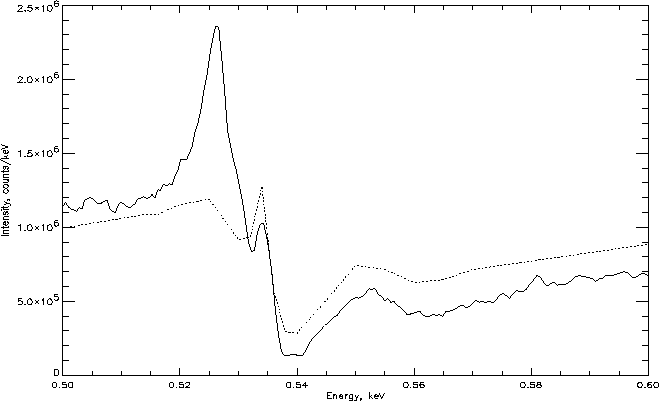
Figure 11: Intensity as function of energy in the vicinity of the oxygen
edge (solid line). Dotted line represents QE plot of the CCD multiplied by
a factor of  to match the scale.
It also means that HIREFS can be used to measure XAFS structure
in the CCD response with very fine energy steps.
An oxygen edge in the HIREFS data is stronger than QE plot predicts.
This can be explained by the fact that some part of the edge comes
from the X-ray source, not entirely being produced be the CCD.
A film of hydrocarbons (most likely, from the vacuum
turbopump) forms on the surface of the target in the X-ray source
contains oxygen, nitrogen and carbon. Emission lines from all
these elements are excited by an electron beam and are
observed in the output spectrum. At the same time, oxygen
contained in the film produces an edge in the spectrum, in
addition to the edge produced by the  in the CCD gate
structure.
To make a mesurement of the oxygen absorption edge of the CCD it is
very important to keep an intensity of oxygen
in the CCD gate
structure.
To make a mesurement of the oxygen absorption edge of the CCD it is
very important to keep an intensity of oxygen  line (525 eV),
emitted by the
source, as
low as possible. For that reason a gold target was used in these
experiments. Gold target does not eliminate
oxygen line entirely, but produces much better results than any other
target we have tried. We were able to reduce oxygen line intensity
to a very low level (see Fig. 11) by keeping an electron
emission current at high levels,
thus increasing target temperature. The achieved line intensity is
about the same as the intensity of the underlying
continuum.
line (525 eV),
emitted by the
source, as
low as possible. For that reason a gold target was used in these
experiments. Gold target does not eliminate
oxygen line entirely, but produces much better results than any other
target we have tried. We were able to reduce oxygen line intensity
to a very low level (see Fig. 11) by keeping an electron
emission current at high levels,
thus increasing target temperature. The achieved line intensity is
about the same as the intensity of the underlying
continuum.
Emission lines of all three light elements seen in the spectrum --
carbon, nitrogen and oxygen have a distinct double-peaked structure.
It is very obvious in the Fig. 2 even
without making functional fits to the spectrum. More detailed quantitative
analysis shows that
oxygen line consists of the wider ( eV) line at 525.0 eV,
and a sharper component ((
eV) line at 525.0 eV,
and a sharper component (( eV) at 526.6 eV.
Nitrogen line appears to be a combination of the peaks at
392 eV (
eV) at 526.6 eV.
Nitrogen line appears to be a combination of the peaks at
392 eV ( eV) and 395 eV (
 eV).
Carbon line structure is even more
complicated, showing peaks at 277 and 279 eV, as well as
few other not so clearly defined peaks at higher energies.
The splitting of the lines is caused
by chemical shifts of the energy levels. An XPS study clearly identifies
at least two types of bonds: C--C and C--O, which explains a complicated
structure of carbon line. It is also very likely that difference between
energy levels of
eV).
Carbon line structure is even more
complicated, showing peaks at 277 and 279 eV, as well as
few other not so clearly defined peaks at higher energies.
The splitting of the lines is caused
by chemical shifts of the energy levels. An XPS study clearly identifies
at least two types of bonds: C--C and C--O, which explains a complicated
structure of carbon line. It is also very likely that difference between
energy levels of  and
and  types of molecular orbitals is responsible for splitting
of the oxygen and nitrogen lines. This is a subject of further
investigation, which can potentially improve an accuracy of the energy
scale of the spectrometer.
types of molecular orbitals is responsible for splitting
of the oxygen and nitrogen lines. This is a subject of further
investigation, which can potentially improve an accuracy of the energy
scale of the spectrometer.